


Preview
Creation Date
10-2-2017
Description
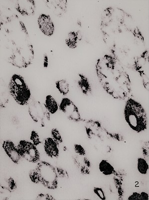
First electron micrograph of a lysosome-rich fraction from rat liver. From Novikoff et al., 1956. J. Biophys. Biochem. Cytol. 2: 179-184
Using only biochemical methods, de Duve came to the conclusion that the new organelle was responsible for hydrolyzing macromolecules in the cell, acting as the “stomach of the cell”. As electron microscopy was simultaneously becoming more developed, it was eventually used to visualize de Duve’s new organelle – the lysosome – in purified cell fragments by Alex Novikoff, a visiting scientist from the Albert Einstein College of Medicine in New York
Keywords
Christian de Duve, Alex Novikoff, electron microscopy, lysosome